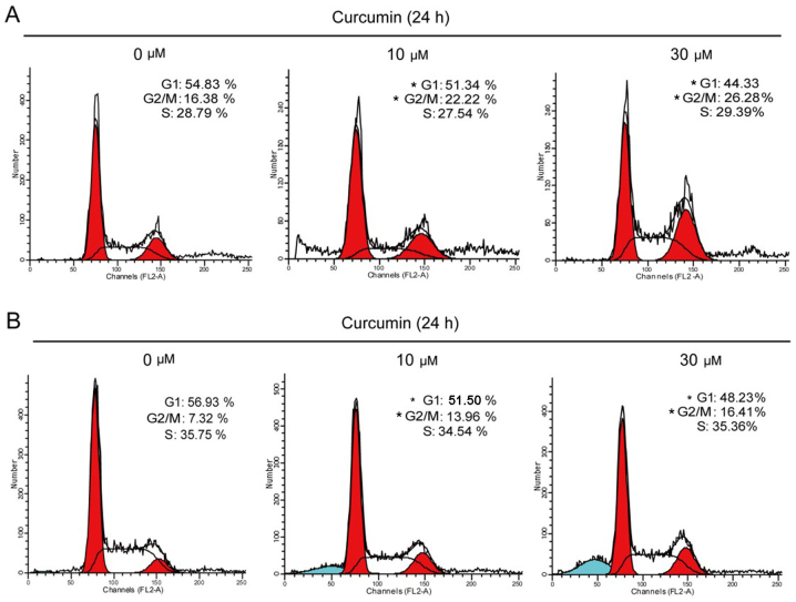
Figure 2.
Curcumin induced G2/M arrest in breast cancer cells. (A) T47D cells were treated with 10 or 30 µM curcumin for 24 h, and the cell cycle was assessed by propidium iodide (PI) staining using flow cytometry. (B) MCF7 cells were treated with 10 M or 30 µM curcumin for 24 h, and the cell cycle was assessed by PI staining using flow cytometry. Data represent mean values from three independent experiments. *P<0.05 compared with the control group by comparing each cell cycle phase.

