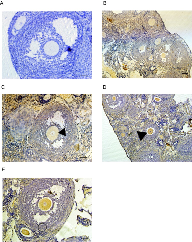
Figure 2.
Ovarian localization of Epac and Rap1 proteins determined by immunohistochemistry. (A) Negative control. The control sections were incubated with PBS only, without the primary antibody and the blue appearance is the result of hematoxylin staining. (B-E) Ovarian sections at various stages of oogenesis with immunostaining for (B and C) Epac and (D and E) Rap1. Different stages of follicles were showed in (B and D) primary follicles, secondary follicles and antral follicles. Arrowheads indicate antral stage follicles in (C and D) and the circle indicates cumulus and granulosa cells in E (scale bar, 20 µm). Rap1, Ras-related protein-1; Epac, exchange proteins directly activated by cyclic adenosine monophosphate.

