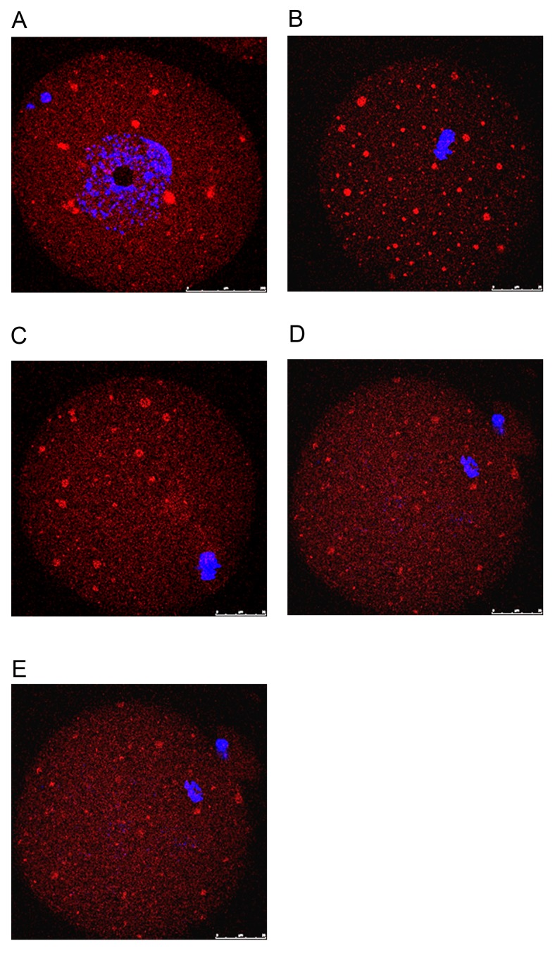
Figure 3.
Localization pattern of Epac during oocyte maturation in vivo and one-cell embryos. (A-E) Different stages of oocytes and embryos were examined for Epac expression (red) using immunostaining. Chromatin was stained with DAPI (blue). (A) Germinal vesicle oocyte. (B) MI oocyte with chromosomes located towards the center of the oocyte. (C) Late MI oocyte. (D) MII oocyte. (E) 1-cell embryos (scale bar, 25 µm). Epac, exchange proteins directly activated by cyclic adenosine monophosphate. M, metaphase.

