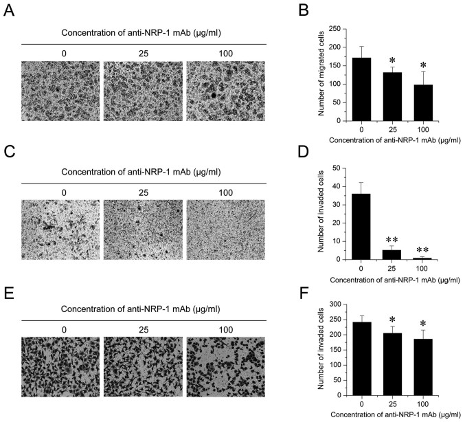
Figure 3.
Anti-NRP-1 mAb suppresses BGC-823 cell migration and invasion. (A and B) A Transwell migration assay was performed to determine the migratory rate of BGC-823 cells treated with different concentrations of anti-NRP-1 mAb (0, 25 and 100 µg/ml) for 12 h. (A) Representative images of migrated cells stained with crystal violet are shown (magnification, ×100). (B) Quantification of migrated cells. (C-F) A Transwell invasion assay was also performed on BGC-823 cells treated with anti-NRP-1 mAb-treated (0, 25 and 100 µg/ml). (C) Representative images of invaded cells across a Matrigel-coated membrane following 12 h treatment with anti-NRP-1 mAb are shown (magnification, ×100). (D) Quantification of invaded cells following 12 h anti-NRP-1 mAb treatment. (E) Representative images of invaded cells following 24 h treatment with anti-NRP-1 mAb are shown (magnification, ×100). (F) Quantification of invaded cells following 24 h anti-NRP-1 mAb treatment. All data are presented as the mean ± standard deviation of three independent experiments. *P<0.05 and **P<0.01 vs. control cells. NRP-1, neuropilin 1; mAb, monoclonal antibody; BGC-823, human gastric cancer cell line; control, untreated BGC-823 cells.

