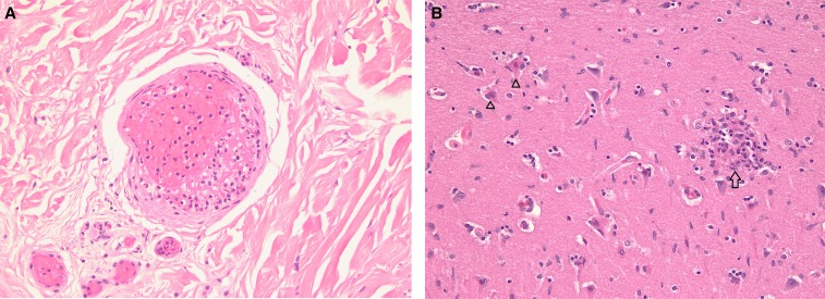
Figure 1.
(A) Dermis with small-vessel thrombosis (hematoxylin and eosin, ×200). (B) Neuronal necrosis (open triangles) with focal perivascular mononuclear cell infiltrate (glial or typhus nodule [open arrow]) (hematoxylin and eosin, ×200). This figure appears in color at www.ajtmh.org.

