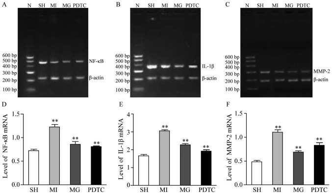
Figure 3.
mRNA expression of NF-κB, IL-1β and MMP-2 in non-infarction zone in different groups. (A-C) Representative gels of the amplimers of (A) NF-κB, (B) IL-1β and (C) MMP-2. (D-F) Quantified expression levels of (D) NF-κB, (E) IL-1β and (F) MMP-2. **P<0.01 for comparison between either two groups. Groups: MI, myocardial infarction model group treated with saline for 28 days; SH, sham; MG, model group treated with MG-132 for 28 days; PDTC, model group treated with pyrrolidine dithiocarbamic acid for 28 days; N, marker; IL, interleukin; MMP, matrix metalloproteinase; NF, nuclear factor.

