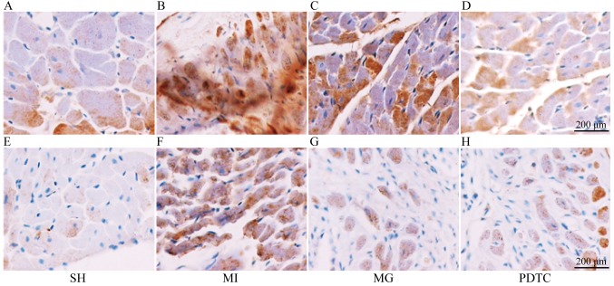
Figure 5.
Immunohistochemical staining for IL-1β and MMP-2. (A-D) Staining for IL-1β in the (A) SH, (B) MI, (C) MG and (D) PDTC groups. (E-H) Staining for MMP-2 in the (E) SH, (F) MI, (G) MG and (H) PDTC groups (scale bar, 200 µm). Groups: MI, myocardial infarction model group treated with saline for 28 days; SH, sham; MG, model group treated with MG-132 for 28 days; PDTC, model group treated with pyrrolidine dithiocarbamic acid for 28 days; IL, interleukin; MMP, matrix metalloproteinase; NF, nuclear factor.

