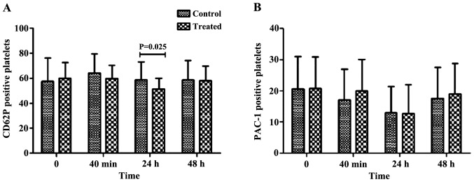
Figure 1.
The percentage of CD62P and PAC-1 detected by flow cytometry. (A) The percentage of CD62P 24 h after PCI in the observation group was significantly lower than that in the control group after PCI (P<0.05). (B) There was no difference in other time-points (P>0.05). There was no statistical difference in the percentage of PAC-1 in the groups at any time-point (P>0.05). CD62P, alpha granule membrane glycoprotein; PCI, percutaneous coronary intervention.

