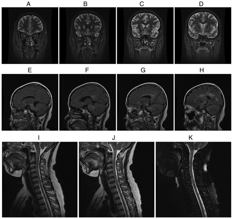
Figure 1.
A female myelin oligodendrocyte glycoprotein antibody(+) and aquaporin-4 antibody(−) neuromyelitis optica-associated ON patient with an onset age of 38 years, disease duration of 16 years and a total of 15 recurrent attacks presented with an attack of ON on both eyes, acute myelitis involving >3 segments, postrema syndrome and acute diencephalic syndrome. (A-D) The brain MRI coronal short-time inversion recovery revealed that the intraorbital segment, canal segment and intracranial segment of optic nerve and optic chiasma became thinner and the signal was increased when compared with an earlier image obtained from patients prior to the present study. (E-H) The brain MRI sagittal T2 fluid-attenuated inversion recovery revealed the abnormal signal in bilateral basal ganglia, thalamus, lateral ventricle, brain stem and corpus callosum. (I-K) Cervical MRI sagittal T2-weighted images revealed the abnormal signal at the medullary-C2 and C5-T1. ON, optic neuritis; MRI, magnetic resonance image.

