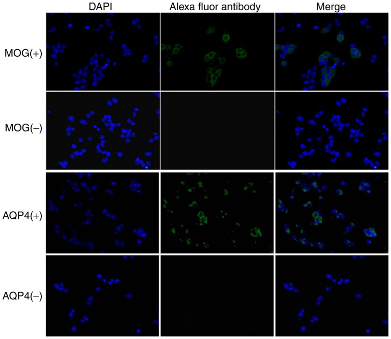
Figure 2.
MOG antibody was detected by a cell-based assay (magnification, ×100). 293 cells were observed under a fluorescence microscope. Blue fluorescence represents nuclei and green fluorescence represents Alexa fluor 488-conjugated MOG antibody. MOG, myelin oligodendrocyte glycoprotein; AQP, aquaporin.

