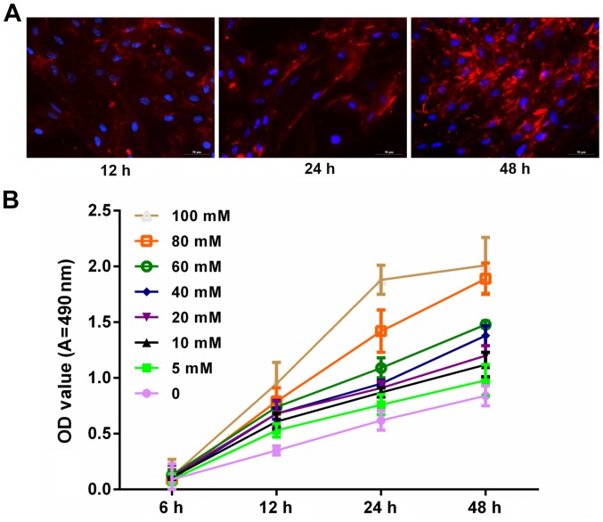
Figure 1.
Glucose promoted podocyte proliferation in a marked time- and dose-dependent manner. (A) Podocytes were cultured at 37°C for 12, 24 and 48 h. The synaptopodin expression was detected using immunofluorescence assay. The cells were visualized under fluorescence microscopy. Blue and red fluorescence represents the nucleus and synaptopodin, respectively. (B) MTT assay was performed to measure the proliferation of podocytes treated with PBS and glucose at 0, 5, 10, 20, 40, 60, 80 and100 mM for 6, 12, 24 and 48 h. OD, optical density; A, absorbance.

