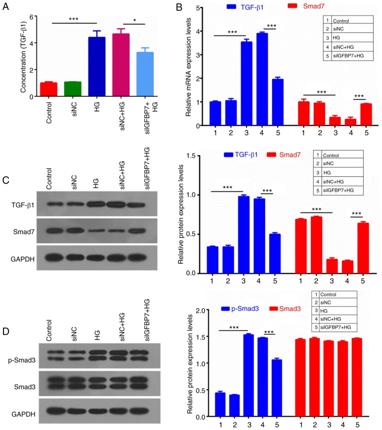
Figure 5.
Silencing IGFBP7 suppressed the TGF-β1/Smad pathway in podocytes induced by HG. Podocytes were treated with PBS (control), siNC, 60 mM glucose (HG), siNC and HG, and siIGFBP7 and HG for 12 h. (A) TGF-β1 concentration was detected using ELISA. (B) The mRNA expression levels of TGF-β1 and Smad7 were analyzed using reverse transcription-quantitative polymerase chain reaction. (C) The protein expression levels of TGF-β1 and Smad7 were analyzed via western blotting. (D) The protein expression levels of p-Smad3 and Smad3 were analyzed via western blotting. *P<0.05; ***P<0.001. IGFBP7, insulin-like growth factor-binding protein 7; TGF-Smad7 were analyzed via western bloSmad, mothers against decapentaplegic homolog; HG, high glucose; siNC, normal control small interfering RNA; siIGFBP7, IGFBP7 small interfering RNA; p, phosphorylated.

