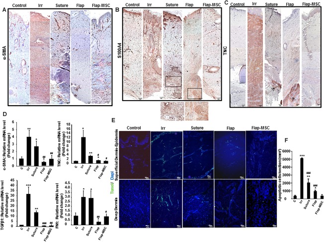
Figure 6.

Bone marrow‐derived mesenchymal stromal cells controlled the fibroblast/myofibroblast accumulation. (A): Representative immunostaining of α‐SMA, an indicator of myofibroblasts showed a staining around the vessels in nonirradiated, suture, flap and flap‐MSC groups and homogenous in the dermis (dark red) in irradiated group. (B): S100a4, a marker of activated fibroblasts, better illustrated in the magnified view in the box (dark red staining), and (C) TNC, an indicator of wound healing throughout the dermis. The TNC staining was intensely homogenous in irradiated group and arrows showed some dark red foci staining in the deep dermis in suture and flap groups. Scale bars: 200 µm. (D): Real‐time expression of wound healing‐related factors, α‐SMA, TNC, and FN1 (fibronectin). (E): TUNEL staining was performed to determine apoptosis. (F): Cellular apoptosis quantification. Scale bars: 100 µm. Results are expressed as means ± SEM. p values were calculated by analysis of variance with Bonferroni correction, *, p < .05; **, p < .01; ***, p < .001 compared with nonirradiated controls; #, p < .05; ##, p < .01; ###, p < .001 compared with irradiated controls. Abbreviations: α‐SMA, alpha smooth muscle actin; MSC, mesenchymal stromal cell; TNC, tenascin C; TUNEL, Terminal deoxynucleotidyl transferase dUTP nick end labeling.
