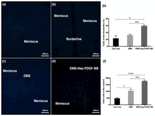
Figure 2.
Cell migration in injured meniscus explants cultured with inserted DMS.
DAPI stained sections of explants cultured for 2 weeks (n=3-6 per group, 10x).
a. Native non-injured meniscus.
b. Injured meniscus cultured without DMS.
c. Injured meniscus cultured with DMS.
d. Injured meniscus cultured with DMS-Hep-PDGF-BB.
e. Graph with numbers of cells at the borderline.
f. Graph with total cell numbers in the explant.
Data represent the mean of 6-8 values from 3 separate experiments.

