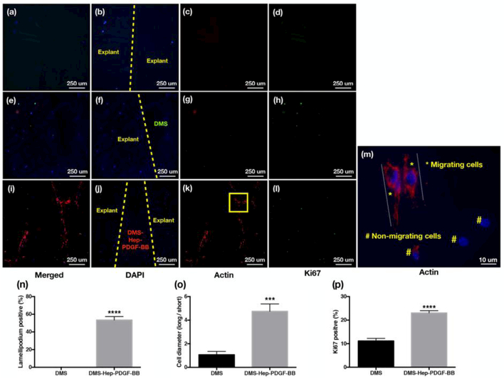
Figure 4.
Migratory and proliferating cells in injured meniscus explants.
DAPI, β–actin and Ki67 stained sections of explants cultured for 2 weeks (n=3-6 per group).
a-d. Injured meniscus cultured without DMS: (a) Merged; (b) DAPI; (c) β–actin; (d) Ki67.
e-h. Injured meniscus cultured with DMS: (e) Merged; (f) DAPI; (g) β–actin (h) Ki67.
i-l. Injured meniscus cultured with DMS-Hep-PDGF-BB: (i) Merged; (j) DAPI; (k) β–actin; (1) Ki67.
m. β–actin staining of injured meniscus cultured with DMS-Hep-PDGF-BB. The elongated actin subunits (lamellipodia) are strongly stained lamella and are indicative of cell migration. Non-migrating cells show negative actin staining or short actin structures. The area marked by a yellow square in panel k is shown at higher magnification in panel m.
n. Graph with % lamellipodium positive cells.
o. Graph with the ratio of cell diameter (long/short diameter).
p. Graph with Ki67 positive cells.
a-l: 20x; m: 60x

