Figure 1.
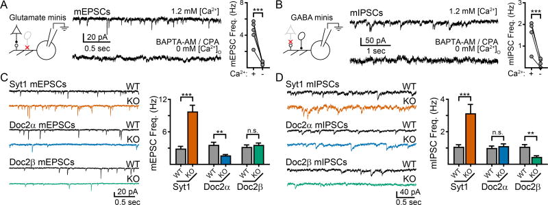
Single KO of syt1, Doc2α, and Doc2β results in different phenotypes for spontaneous excitatory and inhibitory neurotransmission in cultured mouse hippocampal neurons. A. (Left) Schematic of the recording paradigm illustrating that glutamatergic transmission was isolated by inhibiting GABAA-receptors. Glutamatergic neuron: triangle, GABAergic neuron: oval. Representative traces (center) and quantification (right) demonstrating that most AMPA-receptor mediated mEPSCs require the presence of Ca2+. B. Schematic (left), representative traces (center), and quantification (right) demonstrating that GABAA-receptor mediated mIPSCs are also strongly dependent on Ca2+. C. Representative traces (left) and quantification (right) of the effect of single KO of syt1 (orange), Doc2α (blue), and Doc2β (green) on mEPSC frequency compared to WT littermate controls. Syt1 KO resulted in an ‘unclamped’ phenotype and an increase in mEPSC frequency, while Doc2α KO significantly reduced mEPSC frequency. Loss of Doc2β had no effect. D. Same as panel (c), but examining mIPSCs. Syt1 KO again resulted in an ‘unclamped’ phenotype whereas Doc2β KO significantly reduced mIPSC frequency; loss of Doc2α had no effect. ** denotes p < 0.01, *** denotes p < 0.001, n.s. denotes p > 0.05. Bar graphs represent mean ± SEM. See also Supp. Fig. 1 and 2.

