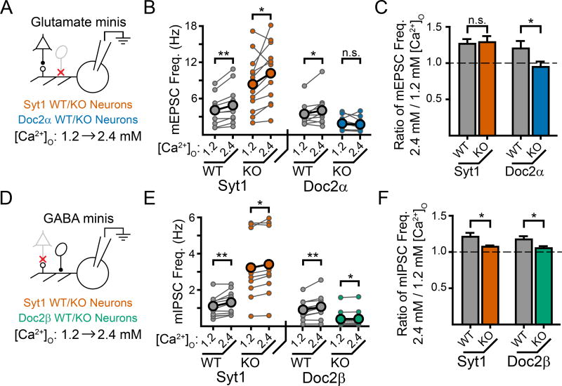
Figure 2.
Doc2α, but not syt1, promotes Ca2+-dependent mEPSCs in a physiological range of [Ca2+]O while both Doc2β and syt1 promote Ca2+-dependent mIPSCs. A. Cartoon schematic of the experimental paradigm. Recordings were made first in 1.2 mM [Ca2+]O before changing the bath solution to 2.4 mM [Ca2+]O. mEPSCs were then recorded again, from the same cell, in 2.4 mM [Ca2+]O. B. Quantification of mEPSC frequency for each recording denoted as a connected pair of smaller circles. The larger circles indicate the average values. C. Quantification of the normalized increase in mEPSC frequency induced by higher [Ca2+]O. Loss of Doc2α abolished this Ca2+-dependent increase while loss of syt1 had no effect. D–F) Experiments were carried out exactly as described in Fig. 2, but examined mIPSCs rather than mEPSCs. Loss of either Doc2β or syt1 reduced, but did not abolish, this Ca2+-dependent increase in mini frequency observed when bath [Ca2+]O was increased from 1.2 to 2.4 mM. * denotes p < 0.05, ** denotes p < 0.01, *** denotes p < 0.001, n.s. denotes p > 0.05. Bar graphs represent mean ± SEM. See also Supp. Fig. 3.

