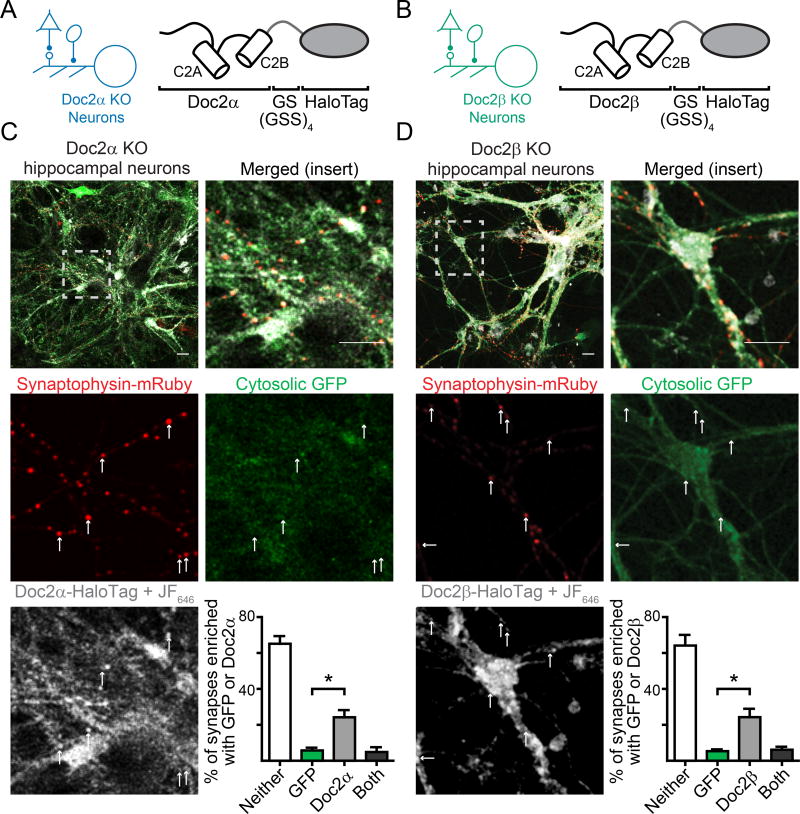
Figure 6.
Doc2 isoforms are enriched at synaptic boutons. A. Schematic demonstrating the fusion of HaloTag to Doc2α and (B) Doc2β. C. Representative live cell confocal images demonstrating that Doc2α-HaloTag (white), expressed at low levels in Doc2α KO neurons, is enriched in synaptic compartments identified by the presence of mRuby-tagged synaptophysin (red). Neurons were co-infected with virus expressing soluble GFP (green), which was not enriched in synapses. The JF646 dye was used because it is significantly brighter than protein fluorophores and fluoresces only when bound to HaloTag proteins Upper-right: quantification of percentage of synaptophysin-positive boutons (synapses) that had punctate enrichment of GFP or Doc2α. D. Similar to C, Doc2β-HaloTag (white) also is enriched in synaptic compartments when expressed in Doc2β KO neurons. * denotes p < 0.05, ** denotes p < 0.01, *** denotes p < 0.001, n.s. denotes p > 0.05. Scale bar represents 20 µm. Bar graphs represent mean ± SEM. See also Supp. Fig. 5 and 6.

