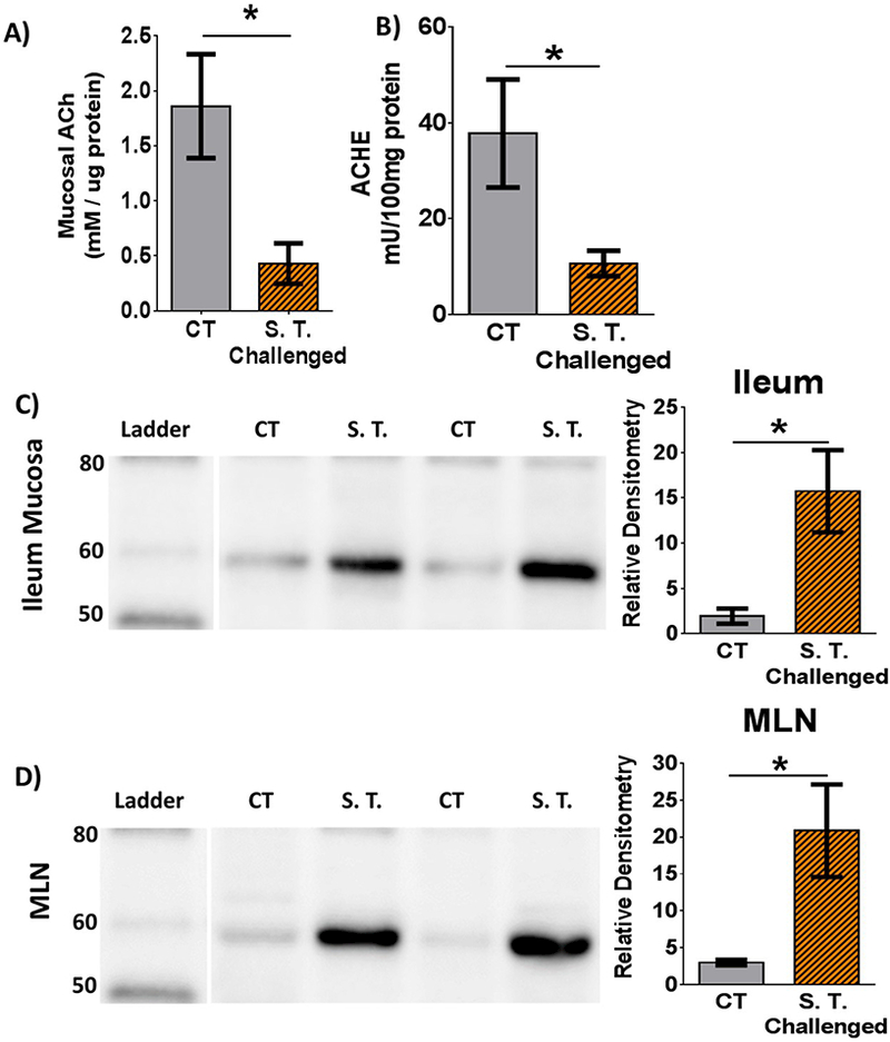
Figure 1. Impact of S. Typhimurium challenge on acetylcholine and cholinergic enzymes in ileum mucosa.

A) Acetylcholine concentration in ileal mucosa. B) Acetylcholinesterase activity ileal mucosa. C) Western blot of ileal mucosa for porcine ChAT. Prominent band is between 60 and 50kDa. Histogram quantifying relative density directly to the right. D) Western blot of mesenteric lymph node (MLN) for porcine ChAT. Respective histograms represent relative density normalized to lane total protein. CT = Control, S.T. = S. Typhimurium. Bars and SEM represented. n = 5 controls and 6 challenged per bar. Student’s t-test compared controls vs S. Typhimurium challenge. * = p<0.05.
