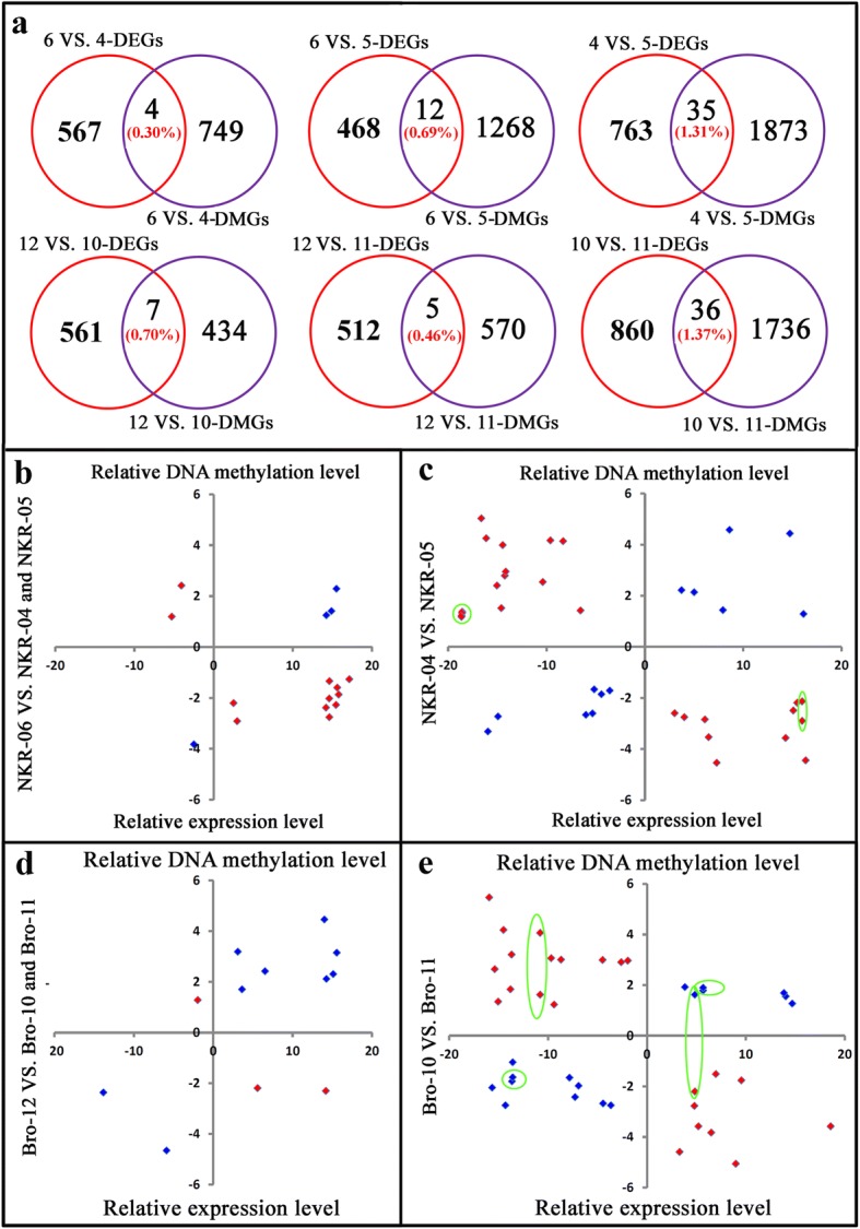
Fig. 9.

Relationship of differential expression levels of genes and their DNA methylation levels. a indicated the Venn diagrams of genes showing differential expression levels and/or differential DNA methylation levels by pairwise comparison analysis. DEGs indicated the differentially expressed genes. DMGs indicated the differentially methylated genes. 4, 5, 6, 10, 11 and 12 indicated NKR-04, NKR-05, NKR-06, Bro-10, Bro-11 and Bro-12, respectively. b, c, d and (e) indicated the relationship of the expression levels of DEGs and their DNA methylation levels. The red colors indicted the expression levels of DEGs were negatively correlated with their DNA methylation levels at the CG and/or CHG sites. The blue colors indicated that the expression levels of DEGs were positively correlated with their DNA methylation levels at the CG and/or CHG sites. X axis indicated the relative expression levels of genes. Y axis indicated the relative DNA methylation levels of genes. The DEGs which showed differentially DNA methylation levels at the CG sites as well as the CHG sites were marked by green circles
