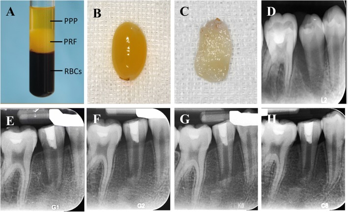
Fig. 1.
a Peripheral blood after centrifugation: red blood cells at the bottom, PRF in the middle, and platelet-poor plasma at the top. b PRF clot. c PRF membrane. d An periapical radiograph of #45 with apical periodontitis in a 12-year-old girl. The case was treated by RET+PRF in #45. e Three-month follow-up periapical radiograph of tooth #45. f Six-month follow-up radiograph. g Nine-month follow-up radiograph. h Twelve-month follow-up radiograph showing complete periapical radiolucency resolution, root apex closure, root elongation and root canal wall thickening

