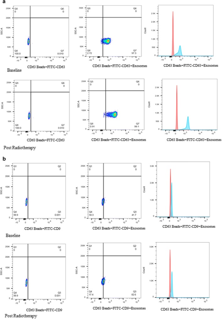
Fig. 4.
Analysis of exosomes by flow cytometry using CD63 coated magnetic beads. Exosomes were first visualized on forward (FSC) versus sidescatter (SSC) plots to gate on the respective exosomes bound to beads population, after gating on singlets. a Typical SSC versus PE-CD63 plots for exosomes isolated from the serum of two donor samples. As comparison, a typical plot for serum derived exosomes from prostate cancer patients using CD63 antibodies at baseline and post-radiation. b Typical SSC versus FITC-A plots for exosomes using CD9 antibodies at baseline and post-radiation. The graph shows increased CD63 representing exosomes compared to CD9 representing surface markers after radiotherapy

