Fig. 3.
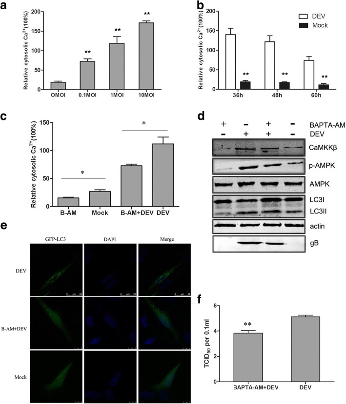
DEV increased cytosolic calcium to activate CaMKKβ and AMPK. a DEF cells were infected with DEV at a MOI of 0.1–10. At 48 hpi,the cells cytosolic Ca2+ were measured based on Fluo 3-AM, a chemical Ca2+ indicator, relative to mock-infected cells. b DEF cells were infected with DEV at a MOI of 1. At 36, 48 and 60 hpi, the cells cytosolic Ca2+ were measured based on Fluo 3-AM relative to mock-infected cells. c DEV-infected cells treated with BAPTA-AM, the cells cytosolic Ca2+ were measured based on Fluo 3-AM relative to control cells. d Whole lysates of cells treated with BAPTA-AM or DEV collected at 36 hpi were subjected to western blot analysis of CaMKKβ, p-AMPK, AMPK,LC3,β-actin and gB. e Representative confocal images of DEV-infected DEF cells with or without BAPTA-AM treatment. GFP-LC3 puncta were analyzed. f Viral titer (TCID50) at 48 hpi. All statistical data are reported as the mean ± SEM of three independent experiments (ns, p > 0.05; *p < 0.05; and **p < 0.01)

