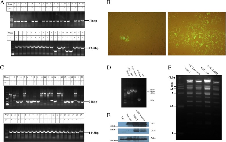
Fig. 3.
Generation and identification of UL41 mutant viruses. a Validation of Gibson-assembled donor plasmids pBlue-eGFP-linker (Top) and pBlue-linker (Bottom) by specific PCR. The sizes of positive PCR products were indicated on right side of each fig. b The donor plasmid pBlue-linker and genomic DNA of ΔUL41 eGFP virus were co-transfected into Vero cells together with or without pLentiCRISPRv1-gRNA. Then, cell culture supernatants were harvested when approximately 80% cytopathic effect was observed. Finally, eGFP protein expression was observed in Vero cells infected with virus supernatants collected from groups transfected with Cas9/gRNA (Left) or not (Right) (Magnification, 200×). c PCR validation of the generated UL41 null virus (Top) and UL41 revertant virus (Bottom). The sizes of positive bands were indicated on right side of each fig. d Identification of each mutant virus by specific PCR. Arrows on the right side indicated the expected sizes of PCR products. e The identification of UL41 protein expression during each virus infection by Western-blot. f BamHI-based RFLP analysis of each PRV strain. The genomic DNA of each virus were prepared and digested respectively with BamHI-HF (NEB) at 37 °C for 3 h. Digested mixtures were then resolved by 0.8% agarose-gel electrophoresis at 60 V for approximately 4 h, and visualized under ultraviolet light

