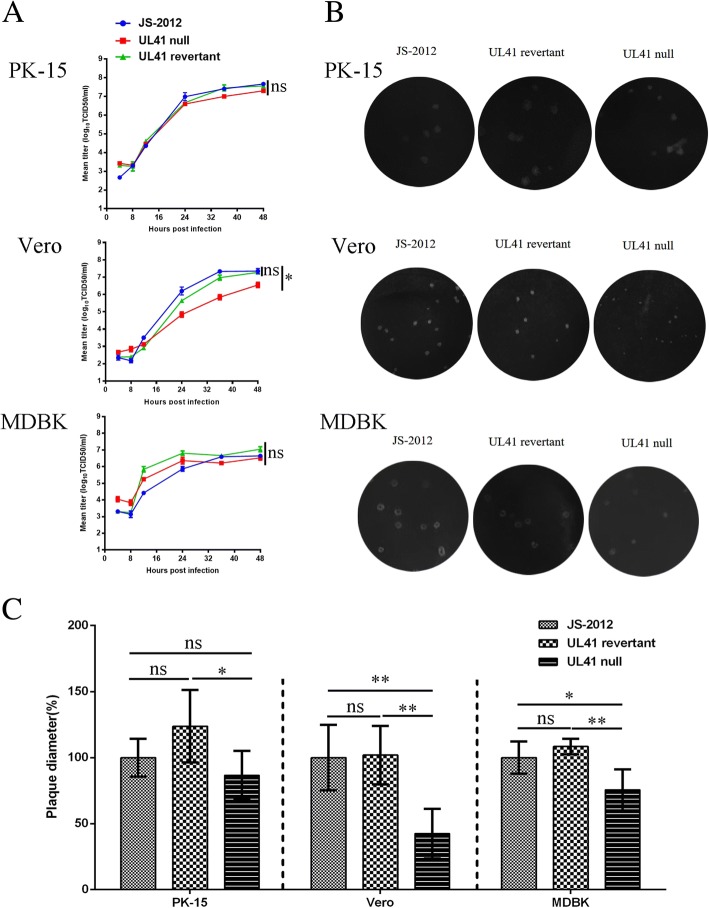
Fig. 4.
Characterization of UL41 mutant virus in vitro. a One-step growth curves. PK-15 (Top), Vero (Middle), and MDBK cells (Bottom) were infected with 1 MOI of each PRV strain. Cell culture supernatants were harvested at 4, 8, 12, 24, 36, and 48 h post infection. Virus titers were determined by TCID50 assay on Vero cells. The mean titers correspond to the averages of two independent experiments. And Two-way ANOVA was used for analyzing the data, *P < 0.05, ns was referred as no significance. b Plaque morphology of JS-2012, UL41 revertant virus and UL41 null virus in PK-15 (Top), Vero (Middle), and MDBK (Bottom) cells cultured at 37 °C for 4 days. c Relative plaque diameters of each virus were calculated and compared to those of PRV JS-2012. Meanwhile the average plaque diameter of PRV JS-2012 in each cells were set as 100%. And the Student’s t-test was used for analyzing the data, *P < 0.05, **P < 0.01, ns was referred as no significance

