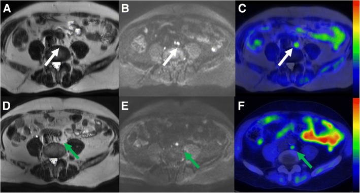
Fig. 6.
Comparison of lymph node staging (N-stage) on pre-transurethral resection (a, b, c) and post-chemotherapy 11C-acetate PET/MRI (d, e, f) in a 66-year-old male (patient number 6 in primary imaging). 8 mm retroaortic lymph node (d - white arrow on T2-weighted image) demonstrated increased diffusion signal restriction (b - b value 800 s/mm2 trace diffusion weighted image), and increased 11C-acetate uptake (c - PET fused with T2-weighted image, SUV is scaled from 0.0 to 2.8) suggestive of lymph node metastasis. On post-chemotherapy 11C-acetate PET/MRI, lymph did not decrease in size and measured 7 mm (d - green arrow on T2-weighted image) with increased diffusion signal (e - b value 800 s/mm2 trace diffusion weighted image) and increased 11C-acetate uptake (f - PET fused with T2-weighted image, SUV is scaled from 0.0 to 2.8). SUVmax values of the lymph node on the pre-transurethral resection (c) and post-chemotherapy 11C-acetate PET/MRI (f) was 1.7, 1.3, respectively. No lymph node metastases were found on extended pelvic lymph node dissection, thus the findings of 11C-acetate PET/MRI were considered as false positive

