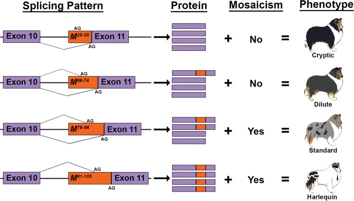
Fig. 8.
Proposed mechanism for merle phenotypic variation. Suggested patterns of splicing are shown for cryptic, dilute, standard, and harlequin merles. The Merle SINE is depicted in orange with oligo(dT) length ranges superscripted. The original and alternative (within SINE) splice acceptor sites are denoted by “AG.” The proportion of wild-type (solid purple) to aberrant (purple and orange) PMEL protein illustrates the proposed frequency of alternative splicing, as it corresponds to oligo(dT) length. Mosaicism reflects whether somatic oligo(dT) contractions were observed in each phenotypic group herein. Together, the rates of alternative splicing and somatic oligo(dT) contractions confer the background coat color intensity and variegation, respectively

