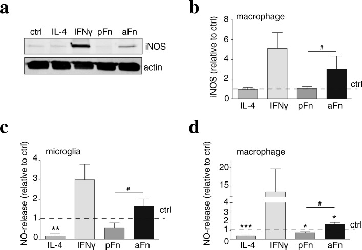
Fig. 5.
Fibronectin aggregates, but not plasma fibronectin, induce iNOS expression by bone marrow-derived macrophages. Microglia (c) and bone marrow-derived macrophages (BMDMs, a, b, d) were left unstimulated (ctrl), cultured on plasma fibronectin (pFn) or fibronectin aggregates (aFn), or treated with interferon-γ (IFNγ) or interleukin-4 (IL-4). Then, the expression of iNOS (a, b) and nitric oxide (NO) levels (c, d), markers for classically activated microglia and BMDMs, were analyzed as described in materials and methods. Note that aFn, but not pFn, increased both iNOS (a, b, n = 4–5), along with an increased NO release (c, d, n = 5–12). Bars represent mean values of each condition relative to control cells (set at 1 for each independent experiment, horizontal line). Error bars show the standard error of the mean. Statistical analyses were performed using the one-sample t test when compared to control (*p < 0.05; **p < 0.01; ***p < 0.001). A student t test was performed to compare pFn with aFn (#p < 0.05)

