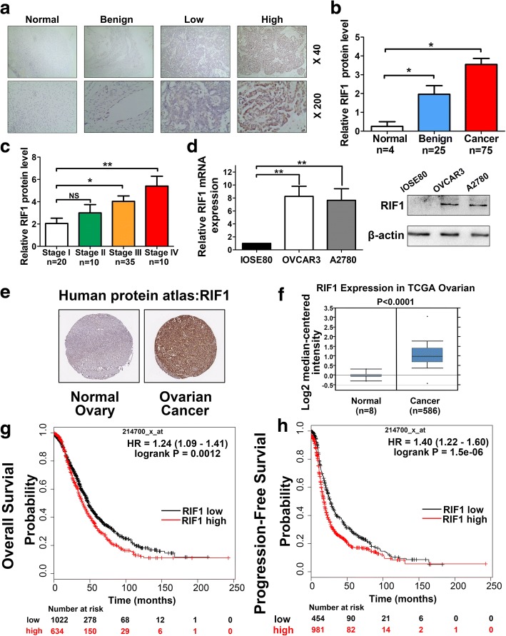
Fig. 1.
Clinical significance of RIF1 in human ovarian cancer patients. a Representative images of immunohistochemistry staining of RIF1 in specimens of EOC, normal and benign ovarian tissues. b Expression level of RIF1 in ovarian cancer progression was detected by IHC. c The expression of RIF1 was examined by IHC in different clinical stages of EOC tissues. d The mRNA and protein expression levels of RIF1 in ovarian epithelial cell line (IOSE80) and two human ovarian cancer cell lines (OVCAR3 and A2780) cell lines were detected by RT-qPCR and Western blot. e RIF1 protein expression in normal ovarian tissue and ovarian cancer specimens. Images were taken from the Human Protein Atlas online database. f RIF1 expression is overexpressed in ovarian cancer tissues compared with normal ovarian tissues. The identified and normalized data were taken from Oncomine database. g, h Kaplan-Meier analysis of overall survival and progression-free survival by low or high RIF1 expression in ovarian cancer patients were performed by using Cox proportional hazard models and follow-up data for 20 years. The data were obtained from Kaplan-Meier plotter database and all plots were analyzed by combing 15 data sets (GSE14764: n = 80, GSE15622: n = 35, GSE18520: n = 63, GSE19829: n = 28, GSE23554: n = 28, GSE26193: n = 107, GSE26712: n = 195, GSE27651: n = 49, GSE30161: n = 58, GSE3149: n = 116, GSE51373: n = 28, GSE63885: n = 101, GSE65986: n = 55, GSE9891: n = 285 and TCGA: n = 565). HR = hazard ratio. * P < 0.05, ** P < 0.01

