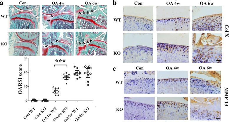
Fig. 2.
a Safranin-O/fast green staining and quantification of the histologic results using the Osteoarthritis Research Society International Cartilage Histopathology Assessment System (OARSI score) indicated articular cartilage damage in all groups. Black arrows show the damaged region of the cartilage. **p < 0.01, compared with the wild-type (WT) osteoarthritis (OA) mice at 4 weeks (4w). Scale bar = 400 μm. Immunohistochemical analysis of b type X collagen (COL X)- and c matrix metalloproteinase 13 (MMP13)-positive chondrocytes (brown) in articular cartilage showed that GPR120 knockout (KO) mice significantly increased the numbers of COL X- and MMP13-positive chondrocytes compared with the WT mice 4 weeks after the OA surgery. The fewest numbers of positive cells could be found in the sham control (Con) in both KO and WT mice, and the highest numbers of COL X- and MMP13-positive cells can be found in OA at 6 weeks (6w) for both KO and WT mice. Scale bar = 50 μm

