
Fig. 1.
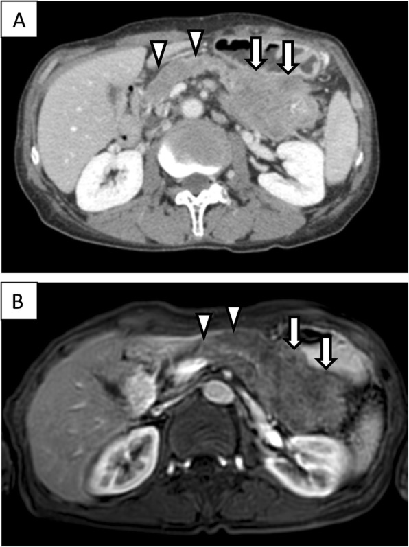
Episode 1: primary pancreatic tumor. Diffuse pancreatic mass in a 67-year-old man. Contrast-enhanced computed tomography (CT, a) scan and magnetic resonance imaging (MRI, b) showed a hypovascular pancreatic mass located mainly in the pancreatic tail (arrows) and involving the entire pancreas (arrow head)