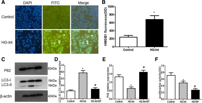
Fig. 4.
HMGB1 mediates intermittent high glucose-induced autophagy. DAPI was used to stain the nuclei. a-b: Immunohistochemistry showed that the normal glucose ARPE-19 cells were mainly expressed as nuclear localization of HMGB1. However, in ARPE-19 cells treated with intermittent high glucose, the proportion of HMGB1 in the cytoplasm was increased. c-d: EP could inhibite intermittent high glucose-induced LC3-II expression. e: EP could increase the expression of p62 protein under conditions of intermittent high glucose. f: Loss of HMGB1 under intermittent high glucose condition resulted in dramatic significantly reduction in proliferative activity than intermittent high glucose and normal glucose(*p < 0.05 vs. control, #p < 0.05 vs. HG)

