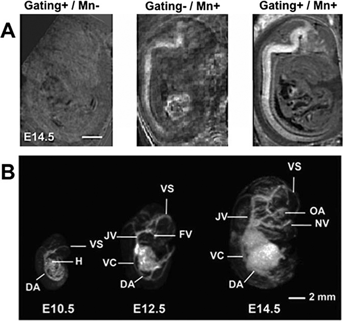
Fig. 2.

In utero Tr-weighted MEMRI and T2*-weighted vascular MRI. (a) Mn-enhancement and respiratory gating work in combination to improve in utero MRI. Images at E14.5 with gating and without Mn (Left panel, Gating+/Mn-), with Mn and without gating (Middle panel, Gating-/Mn+) and with gating and Mn (Right panel, Gating+/Mn+). Scale bar is 1 mm. The images are modified with permission from [5]. (b) 3D maximum intensity projections (MIPs) show the developing vasculature from E10.5 to E14.5. Labels: DA dorsal aorta, FV facial vein, H heart, JV jugular vein, NV nasal vein, OA optic artery, VC vena cava, VS venous sinus. The images are modified with permission from [9]
