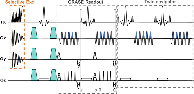
Fig. 4.

A diagram of the 3D DW-GRASE sequence with a spatially selective excitation pulse. The diagram shows the timing of the 2D selective excitation pulse together with the spiral gradient in the x-y plane, the diffusion encoding gradients (represented by turquoise trapezoids), the GRASE readout module, and the twin- navigator echoes. Each GRASE readout module acquires four gradient echoes and one spin echo, and the readout is repeated four times to achieve an acceleration factor of 20 compared to the conventional spin echo sequence. The images are modified with permission from [6]
