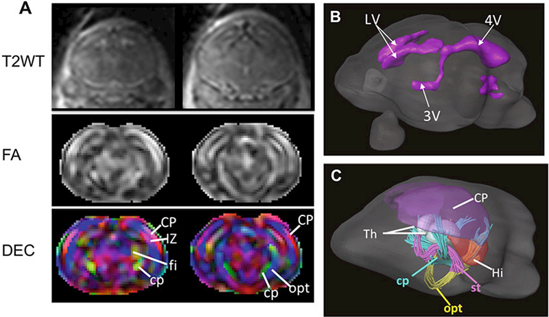
Fig. 6.

In utero MRI of the embryonic mouse brain. (a) Coronal T2-weighted, FA, and directionally encoded colormap (DEC) images of an E17.5 mouse brain. (b) Surface rendering of the ventricules in the E17.5 mouse brain based on the T2-weighted images. (c) rendering of the gray matter structures and early white matter tracts in the E17.5 mouse brain based on the diffusion MRI data. Abbreviations: CP cortical plate, cp cerebral peduncle, IZ intermediate zone, fi fimbria, Hi hippocampus, LV lateral ventricle, opt optical tract, st stria terminalis, Th thalamus, 3Vand 4Vthe third and forth ventricles
