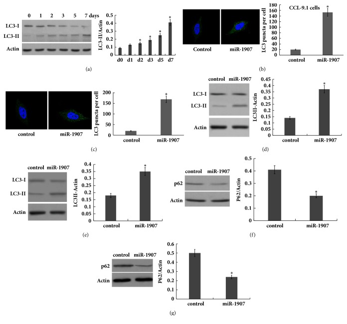
Figure 3.
miR-1907 increases the activation of hepatocyte autophagy. (a) Western blot analysis of LC-3B expression in liver tissues at different timepoints after 2/3 PH. CCL-9.1 cells (b) or HL7702 (c) cells were treated with miR-1907, and the autophagosome formation was visualized by assaying activated green puncta. Punctate staining is indicative for the redistribution of LC3 to autophagosomes. The average number of green puncta per cell with standard deviation for each group is presented. Scale bar, 50 μm. CCL-9.1 cells (d) or HL-7702 (e) cells were treated with miR-1907 and then western blot analysis of LC-3B protein expression to evaluate autophagy. CCL-9.1 cells (f) or HL7702 (g) cells were treated with miR-1907 and then western blot analysis of p62 protein level to evaluate autophagy. ∗p < 0.05.

