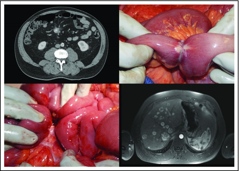
Fig 1.
Top left: A spiculated, partially calcified nodal mesenteric mass seen on computed tomography scan. Top right: Intraoperative image of a primary small bowel neuroendocrine tumor with narrowing of the bowel lumen. Bottom left: Intraoperative image of a mesenteric nodal mass. Bottom right: Contrast-enhanced T1-weighted magnetic resonance imaging in the arterial phase that shows multiple hepatic metastases.

