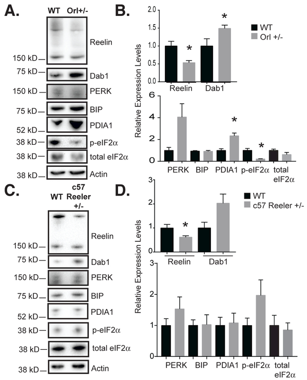
Figure 5. Heterozygous Reeler Orleans (RELN Orl) mice, a model of impaired Reelin secretion, show impaired Reelin signaling and increased PDI expression in the cerebellum.

(A) Western blot of 6-week-old male RELN Orl mice cerebella from wild-type and heterozygous (Orl+/−) mice for Reelin, total Dab1, and ER stress markers. Reelin expression (anti-G10) is reduced and total Dab1 expression (anti-DabH1) is augmented in 6-week-old RELN Orl +/− cerebellum as compared to wild-type (+/+). (B) Quantification of A. Reelin was significantly decreased, while Dab1 was significantly increased in Orl+/− cerebella. PDIA1 was significantly increased in Orl+/− cerebella (n=3, error bars ± SEM, * p< 0.05, t-test). (C) RELN +/− null allele mice cerebellar lysates were analyzed by Western blot for ER stress markers. (D) Quantification of C. Except for reduced Reelin levels in the mutant, no statistically significant differences were seen between the wild-type and RELN +/− null allele cerebella. Dab1 levels tended to be higher in the mutant but this was not significant (n=4-5, error bars ± SEM).
