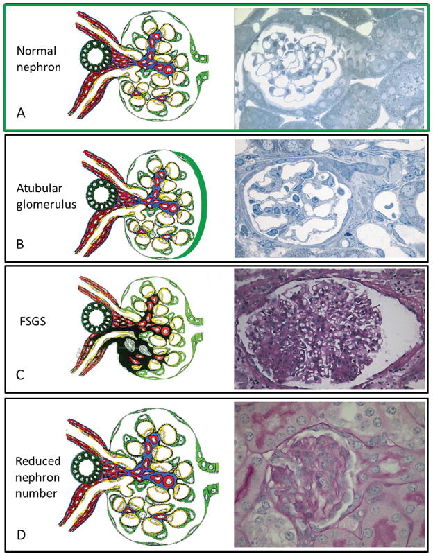
Figure 2.
Structural changes in CKD that have the potential to be detected by CF enhanced MRI technology (diagrams adapted from J. Charles Jennette, MD, www.unckidneycenter.org and micrographs donated by Michael Forbes from Dr. Robert Chevalier’s laboratory). A-Normal structure of a nephron with an intact glomerulotubular junction. B-Atubular glomeruli have been detected in a variety of human diseases and animal models of these diseases. The tubule on the right is closing, as it progresses to a complete disconnection between the glomerulus and tubule. It is felt that glomerulotubular disconnection is an underappreciated mechanism in the progression of chronic kidney disease 68,69. C-FSGS is human renal disease with various lesions that each portends a different clinical course. But due the small sample size obtained on a renal biopsy it is not possible to determine how many glomeruli are sclerotic in a living individual. D–A reduction of nephron number can occur from a congenital etiology or after any renal injury. Although the glomerular structure is normal, an initial and adaptive sign of a nephron reduction is with glomerular hypertrophy. Individual glomerular volume can be determined utilizing CF enhanced MRI.

