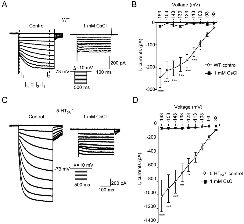
Figure 5. Hyperpolarization-activated inward currents (Ih) in whisker afferent neurons of wild type and 5-HT3−/− mice.
(A) Two sets of sample traces show hyperpolarization-activated inward currents (Ih) recorded from a whisker afferent neuron of a wild type mouse in normal bath solution (control, left panel) and in the bath solution containing 1 mM CsCl (right panel). Inset, hyperpolarization voltage steps. Ih current measurement is indicated in the left panel. (B) Summary data of the Ih currents evoked at different hyperpolarization voltages in normal bath solution (open circles) and in the bath solution containing 1 mM CsCl (solid circles) (n = 8). (C&D) Similar to A&B except the recordings were made from whisker afferent neurons of 5-HT3a−/− mice (n = 9). Data represent the mean ± SEM. *P<0.05, **P<0.01, ***P<0.001, two-way ANOVA with Bonferroni post-hoc tests.

