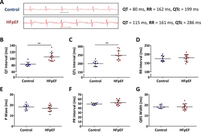
Figure 2. Delayed repolarization in HFpEF rats.

A. Representative ECG showing QT intervals in control and HFpEF rats. B. QT interval was prolonged in HFpEF rats compared to controls. C. QTc interval showed similar trend. D. RR interval was similar in both groups. E, F, G. P wave, PR interval and QRS width were unchanged in both control and HFpEF rats.
