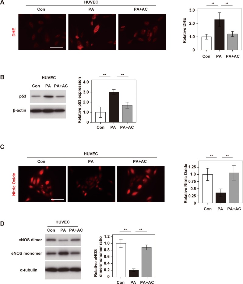
Fig 3. Anthocyanins purified from boysenberry polyphenol protect endothelial cells.
Human umbilical vein endothelial cells (HUVECs) were treated with BSA (Con group), palmitic acid (500 μM) (PA group), or PA (500 μM) + anthocyanins (AC) (10 μg/ml) (PA+AC group). A. DHE staining of HUVECs in Con, PA, and PA+AC groups (Scale bar = 100 μm). The right graph shows the relative fluorescence intensity of DHE (n = 4, 4, and 4). B. Western blot analysis of p53 expression in HUVECs. The right panel displays quantification of p53 relative to the β-actin loading control (n = 3, 3, and 3). C. DAR-4M staining of HUVECs for nitric oxide (Scale bar = 100 μm). The right graph shows the relative fluorescence intensity (n = 4, 4, and 4). D. Western blot analysis of eNOS dimer, eNOS monomer, and α-tubulin expression in HUVECs. The right panel displays quantification of the eNOS dimer/ monomer ratio adjusted for α-tubulin (n = 3, 3, and 3). Data were analyzed by 2-way ANOVA, followed by Tukey’s multiple comparison test. *P < 0.05; **P < 0.01. Values represent the mean ± SEM.

