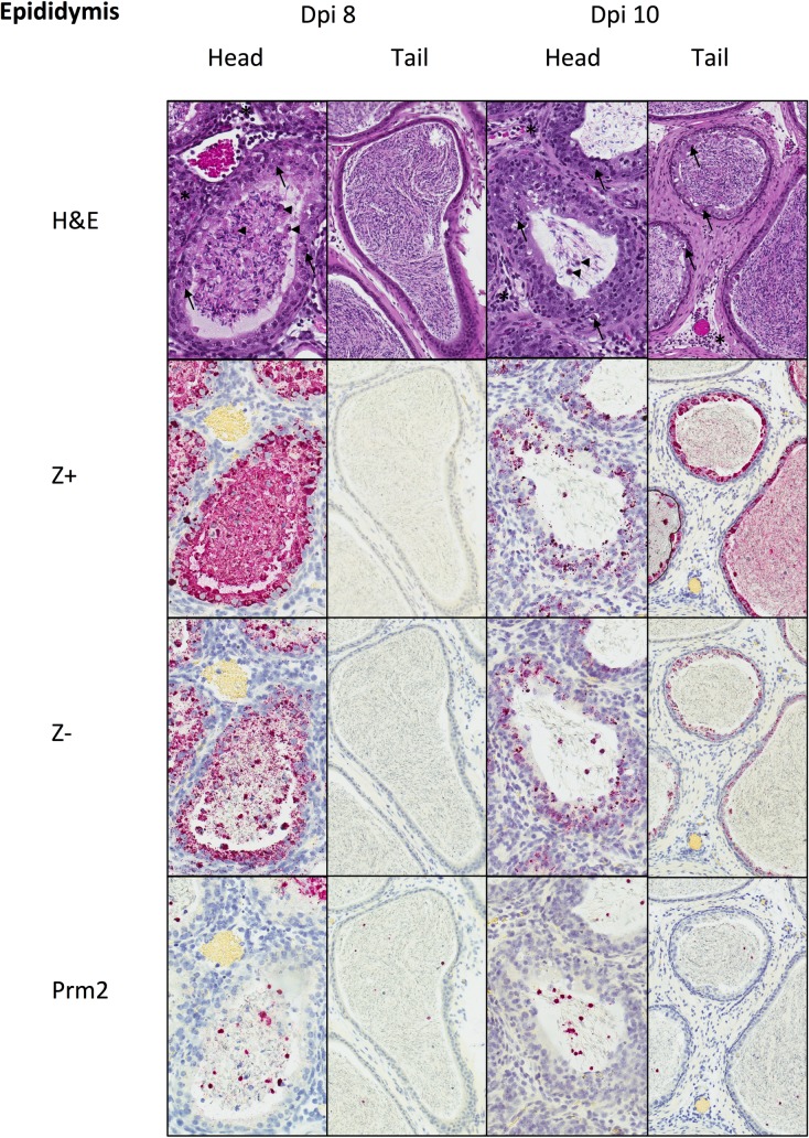
Fig 3. Histopathology and tissue ISH in PRVABC59-inoculated mouse epididymis at dpi 8 and 10.
At dpi 8, the head of the epididymis shows epithelial necrosis (arrows) and mild interstitial inflammation (*). Tubular lumens contain spermatozoa and degenerating round cells (arrowheads). The tail of the epididymis showed no epithelial changes and contained mature spermatozoa. RNA genomic RNA (Z+) and replicative intermediates (Z-) were present within epididymal epithelium and luminal immature spermatozoa and round cells in the head of the epididymis, but not in epithelium or mature spermatozoa in the tail of the epididymis. Many intraluminal round cells were Prm2+ spermatids. At dpi 10, epithelial necrosis and inflammation were similar in the head of the epididymis as seen at dpi 8, and these changes were also seen in the tail of the epididymis. ZIKV genomic RNA (Z+) was decreased in the epithelium and luminal cells of the head of the epididymis, and was also extensively present in the epithelium and spermatozoa of the tail of the epididymis. ZIKV replicative intermediates (Z-) were also present in the epididymal epithelium and in luminal round cells of both the head and the tail, but not in mature spermatozoa of the tail. Many intraluminal round cells were Prm2+ spermatids. H&E and ISH with hematoxylin counterstain of nuclei, Original magnifications: 400x (head), 200X (tail).

