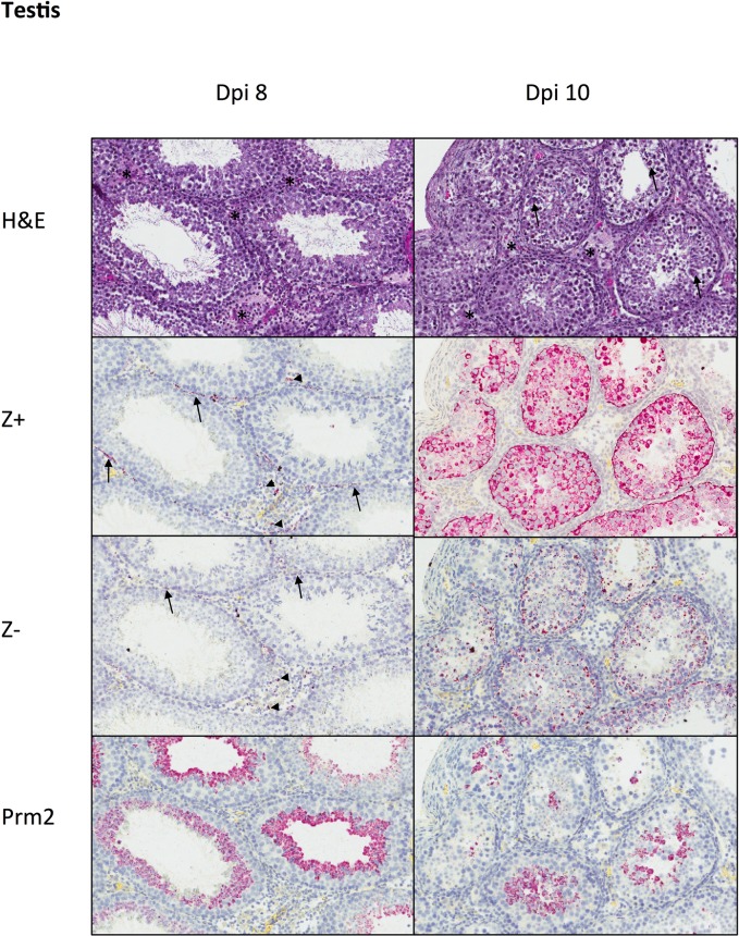
Fig 4. Histopathology and tissue ISH in PRVABC59-inoculated mouse testis at dpi 8 and 10.
At dpi 8, testis showed mild interstitial inflammation (*), without alterations in seminiferous epithelium. ZIKV genomic RNA (Z+) and replicative intermediates (Z-) were localized to scattered interstitial leukocytes (arrowheads) and peritubular myoid cells (arrows). Prm2+ staining showed round spermatids along the luminal surface of seminiferous tubules. At dpi 10, mild interstitial inflammation (*) was accompanied by patchy degeneration of seminiferous epithelium (arrows). Zika virus genomic RNA (Z+) and replicative intermediates (Z-) were present within all layers of seminiferous epithelium, and there was a reduction in the number of Prm2+ round spermatids compared to dpi 8. H&E and ISH with hematoxylin counterstain of nuclei, Original magnifications: 200X.

