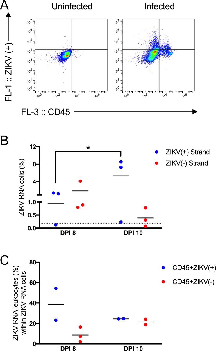
Fig 6. Testicular leukocytes contained replicating ZIKV RNA.
(A) Representative dot plots showing testicular cells stained with ZIKV RNA (+) strand probes (y-axis) and anti-CD45 antibody from an uninfected or infected mouse (x-axis). (B) Percentage of ZIKV RNA (+) or (-) strand cells. On dpi 8, none of the cells stained for ZIKV RNA (-) strand above background. On dpi 10, two of the three mice contained cells in the epididymal lumen that stained for ZIKV RNA (-) strand above background. (C) Percentage of ZIKV RNA leukocytes within ZIKV RNA cells. Each symbol represents one mouse. P-values were determined by 2way ANOVA. *, p<0.05.

