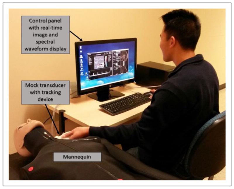
Figure 1.

Photograph of the duplex ultrasound simulator in use. As the examiner moves the mock transducer over the mannequin, a 2-dimensional (2D) B-mode image derived from a saved 3-dimensional (3D) data set is displayed. A Doppler spectral waveform display is generated in real time from the velocity database as the examiner positions the Doppler sample volume and adjusts the control panel settings.
