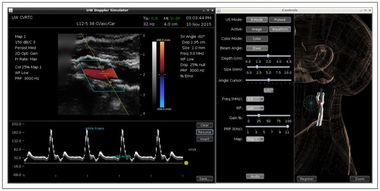
Figure 6.
Measurement of peak systolic and end-diastolic velocities in a simulated distal common carotid artery stenosis. Left panel: The Doppler spectral waveform has been acquired at a beam angle of 60°, and cursors have been placed on the displayed waveform. The measured velocities are 204.7 cm/s peak systolic and 39.6 cm/s end diastolic. Right panel: Ultrasound display mode and Doppler controls and the 3-dimensional (3D) display.

