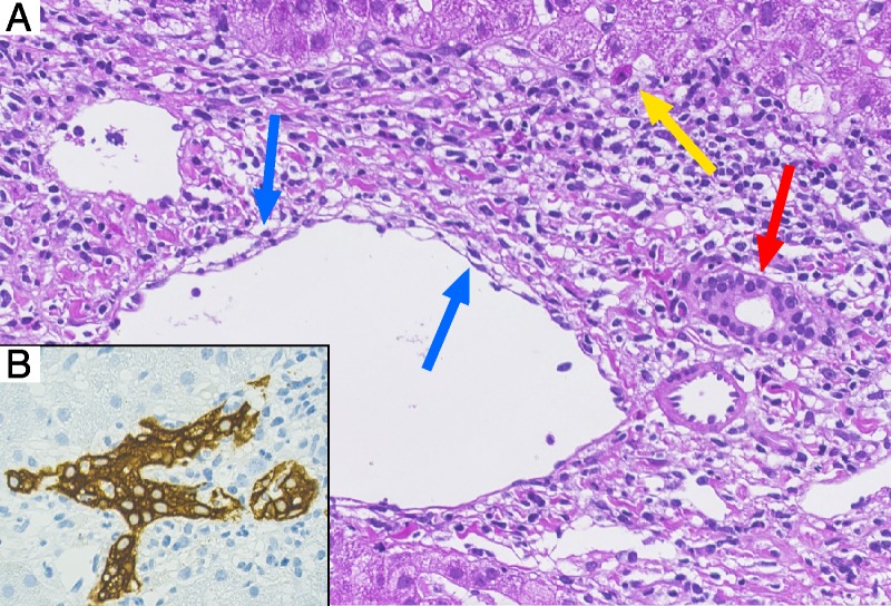
FIGURE 2.

Liver biopsy 16 days after nivolumab. A, HE staining illustrating prominent portal mixed inflammation with interface activity and isolated hepatocyte necrosis (yellow arrow), cytoplasmatic vacuolization of the duct epithelium consistent with bile duct damage (red arrow) and subendothelial lymphocytic inflammation with lifting up of the endothelium compatible with endothelitis (blue arrow); 400×. B, Cytokeratin-7 immunohistochemical staining highlighting the influx of inflammatory cells in the ductal epithelium associated with dysmorphic changes of the bile duct (magnification, 400×).
