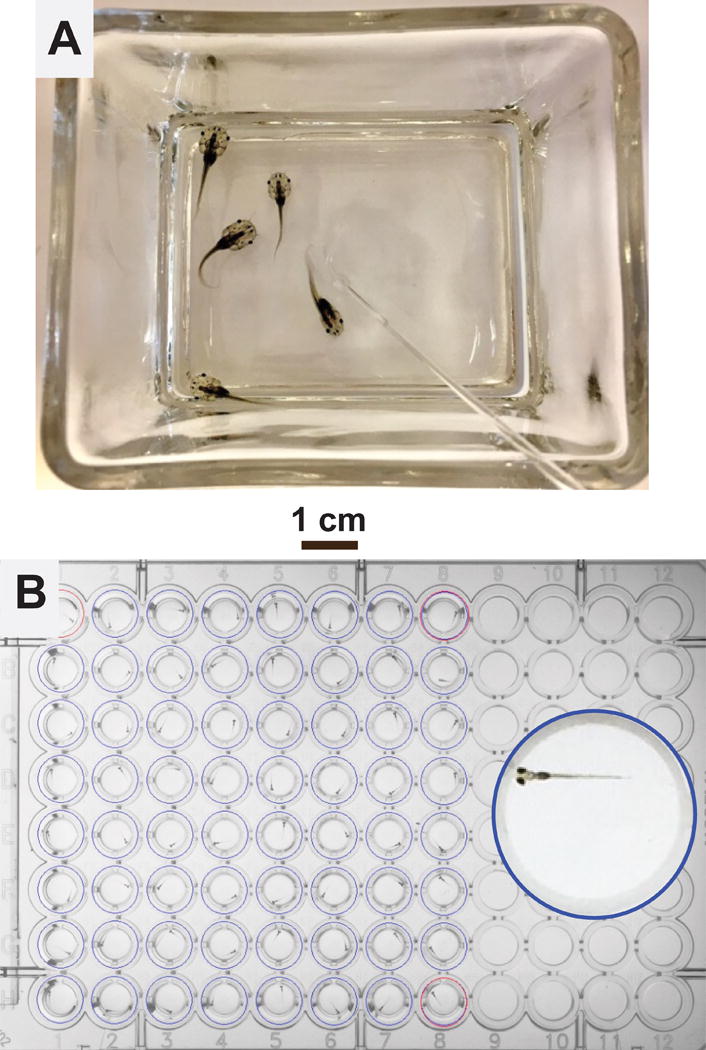
Figure 1. Tadpoles vs. Zebrafish Larvae.

A: A glass container holding 5 pre-limb bud stage Xenopus tadpoles in 100 mL water. The polished glass rod, visible in the lower right quadrant of the photograph, is used to manually turn the animals during loss-of-righting reflexes tests.
B: A 96-well plate loaded with 64 zebrafish larvae, 1 larva per well in 0.2 mL E3 buffer each. The inset shows a magnified view of one well containing a 7 dpf larva.
