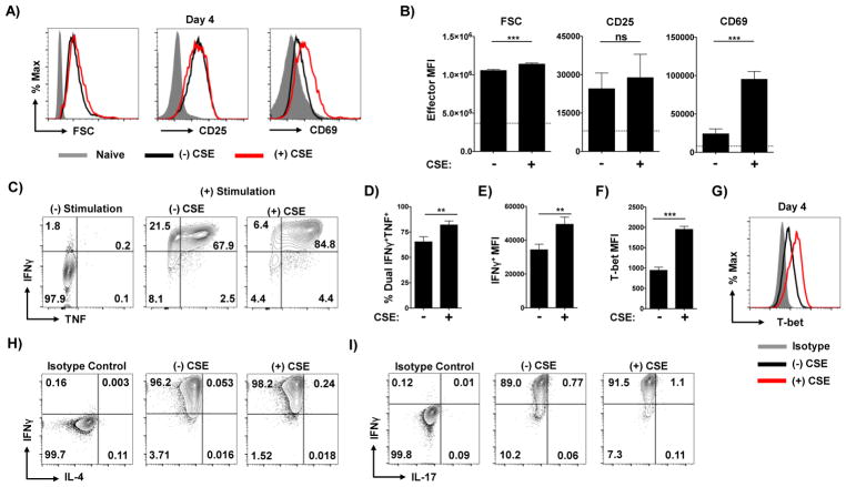
Figure 2. Exposure to CSE during priming enhances Th1 polarization.
Naïve CD4 T cells were stimulated with APC and peptide under Th1 conditions in the presence or absence of CSE. Effector cells were harvested on day 4 of culture and analyzed for forward scatter (FSC), CD25, and CD69 with (A) representative staining and (B) mean fluorescence intensity (MFI) analysis from triplicate cultures shown. Values from naïve CD4 T cells are shown as grey histograms in (A) and as dotted line in (B). Effector cells were assessed for their capacity to produce of IFNγ and TNF by intracellular cytokine staining. Shown is (C) representative staining in the absence or presence of stimulation by PMA and Ionomycin by effectors generated in the presence or absence of CSE, as well as (D) the frequency of cells from each condition capable of co-producing TNF and IFNγ and (E) the MFI of IFNγ+ cells from triplicate wells (one of 4 experiments). Expression of T-bet in effectors was determined by intracellular staining with MFI analysis and representative staining shown (F and G). Representative staining of re-stimulated cells for production of IL-4 (H) and IL-17 (I) (one of 2 experiments).

